Technical Surgery Details
If you are anything like me and you want detailed answers to all the questions… this post is for you. So just hang with me because I’m going to do a little review for all the newbies joining us. But I’ll include pictures with lots of detail to bring everyone up to speed.
So let’s start with What is Chiari Malformation? Info Source
Chiari malformation (kee-AH-ree mal-for-MAY-shun) is a structural abnormality at the back of the brain and skull. Normally, a large hole in the base of the skull accommodates the connection between the brain and spinal cord. This connection point is surrounded by fluid that can move freely between the head and spine. In someone with a Chiari malformation, the back of the brain (the cerebellum), is pushed down through this opening, creating pressure on the spinal cord and restricting that fluid movement between the head and spine. That pressure leads to a wide variety of symptoms.
Those symptoms may include:
Headache that gets worse with exertion, including exercise, coughing, sneezing, or laughing. The pain may also be experienced with certain movements, such as bending forward. (Henley was having these symptoms a lot before her second surgery)
Neck pain, specifically at the base of the neck or between the shoulder blades.
Dizziness (Henley had a lot of this around age 7 before her second surgery)
Tingling or numbness, usually in the hands (At age 7 Henley had this in hands and in her feet)
Unsteady gait (unbalanced, clumsy, she basically looked drunk while walking a straight line. Henley is also currently having trouble with some motor planning things like walking down stairs, getting on escalators and overall coordination)
Loss of fine motor skills (handwriting, using scissors etc: Current issue)
Choking on liquids (Henley struggled with this at Age 2 & again at age 7)
Spine deformity (scoliosis)
Speech Issues (Current Symptom)
Difficulty swallowing (Henley had increased swallowing issues before her second surgery, she would chew on food and eventually just spit it out because it was hard to swallow)
Gagging or vomiting (Just about every single feeding from 18mths until her surgery involved gagging or vomiting)
Developmental delays
Failure to gain weight (Henley was considered “Failure to thrive” when she was a baby because she couldn’t gain weight no matter how much we fed her)
Other symptoms that Henley had that were not official documented “Chiari Symptoms” were:
Seizures (we believe this was due to the amount of compression on her brain stem. This completely resolved after her second surgery and we have not seen one since
Cognitive “cloudiness” After Henley’s second surgery it was like a curtain was pulled back and she was able to mentally process things so much faster. She had more words to say, spoke clearer and faster and seemed to have a lot more energy.
What does the Cerebellum Control in the Brain? Info Source
The cerebellum has several functions relating to movement and coordination, including:
Maintaining balance: The cerebellum has special sensors that detect shifts in balance and movement. It sends signals for the body to adjust and move.
Coordinating movement: Most body movements require the coordination of multiple muscle groups. The cerebellum times muscle actions so that the body can move smoothly.
Vision: The cerebellum coordinates eye movements.
Motor learning: The cerebellum helps the body to learn movements that require practice and fine-tuning. For example, the cerebellum plays a role in learning to ride a bicycle or play a musical instrument.
Other functions: Researchers believe the cerebellum has some role in thinking, including processing language and mood. However, findings on these functions are yet to receive full exploration.
What does your brainstem do? Info Source
Your brainstem sends messages between your brain and other parts of your body. Your brainstem helps coordinate the messages that regulate:
Balance.
Breathing.
Facial sensations.
Heart rhythms.
Swallowing.
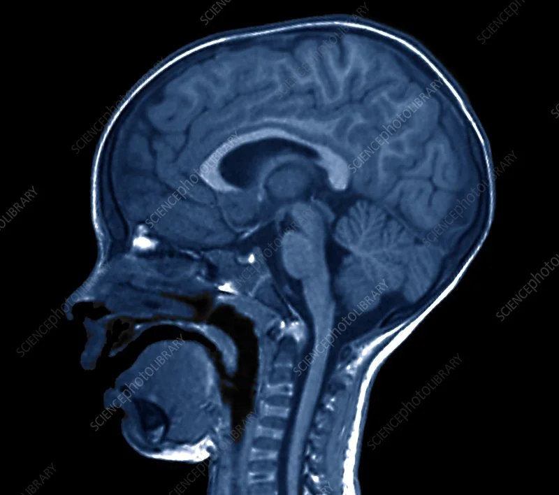
So now you know why those things are super important, the rest of this will make more sense. This first picture is a NORMAL brain. Source: Science Photo Library
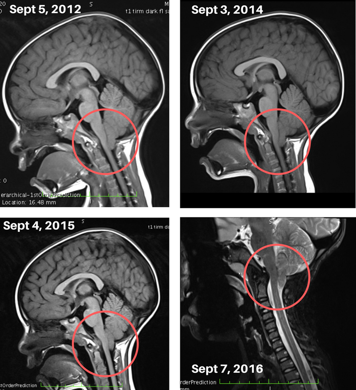
Okay so let me get into some imaging to show you what Henley’s brain actually looks like. Below are pictures of Henley’s brain AFTER her first surgery in 2012 when she was 2 years old. Each year I went in to our local neurosurgeon telling him that while yes, she had stopped vomiting every single day and a few things had certainly improved, we still felt like there were some issues but weren’t sure what to attribute to. We went year after year with basically the same response. “These things aren’t Chiari related”. It made me feel defeated and like I was crazy.
In 2016, I knew in my gut that something was not right. So I set out for a second opinion and wanted to find the most knowledgable person I could in the world of Chiari to talk to. We met with tons of professionals, had sleep studies, swallow studies, speech and occupational therapy evaluations, MRIs, CT scans, X Rays and ended up in New York sitting in front of Dr. Greenfield in December of 2016. All of our speculations of this actually being “Chiari related” were validated and surgery was scheduled for a few months later. Here is a before and after video of what Henley’s brain looked like pre-and post op in March 2017.
Here is one of Dr. G’s PA’s showing the before and after images of Henley’s brain from March 2017 - June 2018
Here is Dr. G explaining what he sees on her MRI from Oct 2022
So what is the surgery going to look like this time? Basically if you watch those two videos back to back you can see how great everything looked in Henley’s brain in June of 2018 (1 year post op) so the plan is to repeat the same surgery and get things back to the way they were then and hopefully they should stay this way for the long haul.
Why was there a change from June 2018 to October 2022? Well the simplest answer is that she has doubled in size since she was 7 years old and with that comes brain and skull growth. With Chiari, that is the issue, the brain needs more room sometimes than the skull will allow, therefore it causes compression of the brain up against the brainstem which causes all kinds of symptoms and that is what we are dealing with currently.
He talked about ossification of the dura tissue in the video, what does that mean? Simply: The definition of ossification, or osteogenesis, is the process of bone formation.
What is the pericranium? The membrane (or periosteum) which covers the outer surface of the skull. This is where they harvest tissue from Henley’s own head to create a “patch” that will be used to help create a larger space for her brain. Sometimes they use cadaver or bovine tissue, but being able to use your own tissue (analogous tissue) is ideal because you have less risk of rejection. They will be using this tissue again but Dr Greengfield feels much more certain that because of her age that hopefully the tissue is less osteogenic (full of stem cells that are set on creating bone).
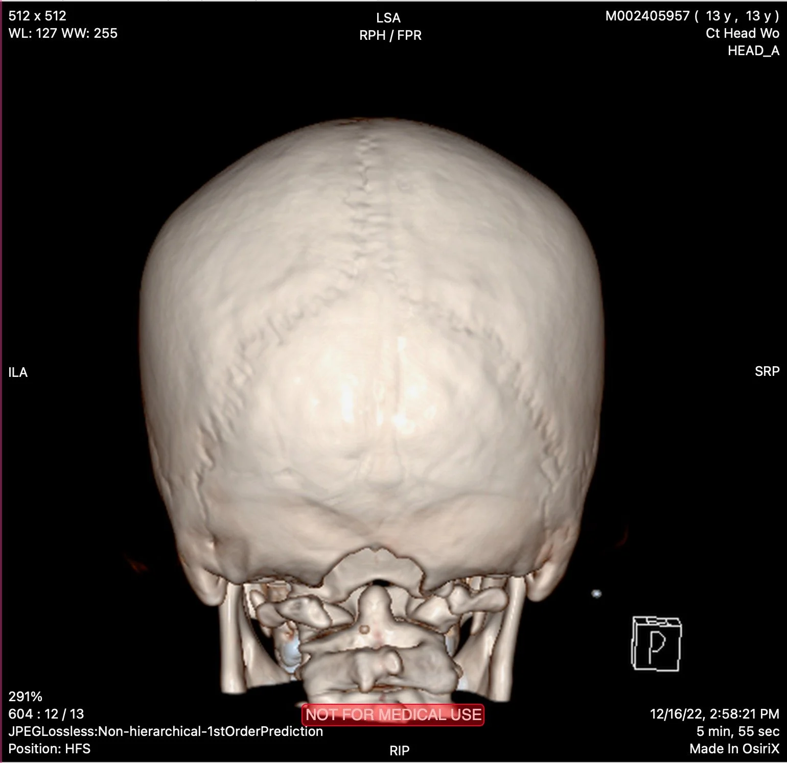
Just because I have an actual picture of this, here is a picture of Henley’s skull in 3d. Kind of wild, I know. But if you look, you can see the half moon shape taken out of the bone at the base of her skull. This is where they have removed bone and will remove a little more bone this go round to allow more space back there. This hole and the fact that Henley is missing part of her C1 vertebrae is the reason she can’t do things like trampolines and jerky rollercoasters. Not a great idea if you are missing skull bone in that area that provides protection around your brain. Keep in mind, you have a layer of muscle over this area and then she has the “patch” that will be sewn into the covering around her brain. So it’s not like her brain is just fully exposed here.
All of these things to say, they will be repeating the same surgery as last time and going in to help relieve the pressure being put on her brain stem by her cramped cerebellum. It is possible that once he is in there he may choose to cauterize some of the cerebellum tissue if it does not look healthy but we won’t know that until Wednesday once they are in there.


